mozanunal.com / Posts / Electrical Impedance Tomography: How It Works and Where It Is Used
Electrical Impedance Tomography: How It Works and Where It Is Used
· 9 min read
Table of Contents
Hello everyone! Today’s topic is Electrical Impedance Tomography (EIT), a cheap, portable, and radiation free imaging modality that builds images from the differences in electrical impedance between tissues. I will first explain how EIT works, then go through some of its clinical applications and research projects, and finally talk about where the field might be heading.
What is EIT?
The name says most of it. Electrical impedance is a measure of how much a material opposes an alternating current. Tomography comes from the Greek words tomos (slice, section) and graphein (to write, to image). Put together, EIT is a technique that estimates tissue boundaries and abnormalities inside the body by measuring impedance from electrodes placed on the skin.
A Short History
- 1950s: Nyboer and colleagues measured blood volume changes in the arm using four electrodes. This technique is known as impedance plethysmography.
- 1962: Thomasset discovered the relation between bioimpedance and total body water.
- 1966: Kubicek and colleagues built the first clinical device that could measure the stroke volume of the heart noninvasively. It was tested on NASA astronauts.
- 1970: Nyboer extended the theory by using the resistivity of blood to measure variations in blood flow.
- 1978: Henderson and Webster turned EIT into a medical imaging system.
- 1980: Settle and colleagues developed methods for nutritional assessment based on whole body and fluid compartment impedance.
- 1980: Alberto P. Calderón laid the mathematical foundation of the inverse problem behind EIT, later extended by Uhlmann (1999).
- 1984: Barber and Brown developed the first practical medical EIT system.
- 2011: The first commercial medical EIT device was introduced for monitoring lung function in intensive care patients [1].
How EIT Works
Tissue Impedance
Different tissues have quite different impedance values, as the tables below show. To measure them, a small and harmless AC current is injected into the body. AC is used because impedance is a complex quantity: its real part is the resistance and its imaginary part is the reactance. Since the reactance depends on frequency, the way a tissue responds to different excitation frequencies is itself another feature that helps distinguish one tissue from another.
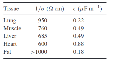
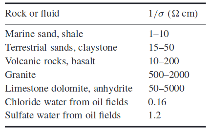 Impedance of different materials and tissues at 10 kHz [2]
Geometry
The geometry of EIT is very different from X-ray CT. In CT, almost all X-ray photons travel to the detector along straight lines, which makes the forward model simple. In EIT, the current does not flow in straight lines. It spreads through the whole volume along curved paths, as in the figure below. Notice that the current lines become sparser toward the center of the object. This is why the spatial resolution of EIT drops in the middle of the imaged region.
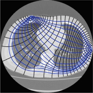 Geometry of impedance measurements [3]
Measurement Strategies
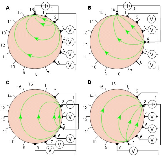
Which electrodes inject current and which ones measure voltage is a design choice. Changing this pattern trades off spatial resolution and SNR in different parts of the image, so different body regions call for different strategies.
Neighboring method. Current is injected through a pair of adjacent electrodes and the voltage is measured across all the other adjacent pairs. Then the current source moves to the next pair, and the process repeats until it has gone around the whole circle. This method gives better resolution near the boundary.
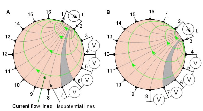 The neighboring method [3]
Cross method. Current is injected between electrodes on crossing sides and the voltages are again read from the remaining electrode combinations. The injection pair is then rotated around the circle.
 The cross method [3]
Opposite method. Current is injected through diametrically opposite electrodes, the voltages are measured, and the injection pair is rotated around the circle. Because the current passes straight through the body, this method gives better resolution in the center.
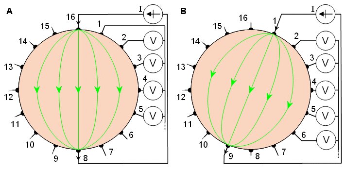 The opposite method [3]
Image Reconstruction
Reconstruction in EIT is much harder than in CT, because the inverse problem is nonlinear and ill posed: very different conductivity distributions can produce almost the same boundary voltages. A classic and fast approach is the Barber-Brown back projection, which is conceptually similar to filtered back projection in CT, except that the measurements are projected back along equipotential lines instead of straight lines. More modern systems use regularized, linearized solvers built on a finite element model of the body. As an example, the figure below shows how lung imaging for volume measurement can be modelled.
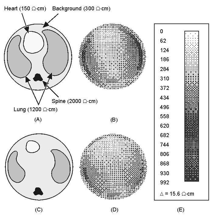 Example reconstruction [1]
Spatial Resolution
Spatial resolution is one of the most critical limitations of EIT, because it is bounded by how many electrodes you can place over the region of interest. Commercial systems typically use 16 electrodes, and some research systems use 32. Still, a few studies have pushed resolution much further.
One impressive example images evoked neural activity in the brain of a rat using a planar electrode array placed directly on the cortex. The array packs many electrodes into an area of about 6 mm x 5 mm and reaches a spatial resolution of around 200 µm [4].
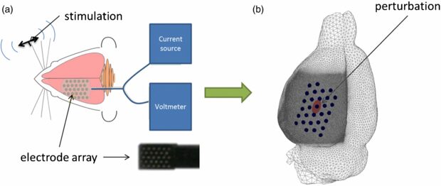 Electrical impedance tomography using intracranial planar arrays [4]
Since the electrodes lie on a plane, resolution depends on depth: the deeper you go into the brain, the coarser the reconstruction becomes. The mesh used in the study reflects this. The finest elements are kept directly under the array down to a certain depth, and then the element size grows linearly toward the opposite side.
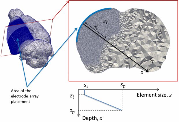 Mesh element size grows with depth below the planar array [4]
Advantages and Disadvantages
| Advantages | Disadvantages |
|---|---|
| Non-invasive | Rarely used and not well known |
| Non-ionizing | Electrode contact problems |
| Compact and portable | Low spatial resolution |
| Inexpensive | |
| High time resolution |
Applications
Lung Monitoring
Lung monitoring was the first commercial clinical application of EIT, and it is a natural fit. Patients in intensive care need continuous monitoring, and EIT devices are small enough to stay at the bedside. The electrodes are attached with a belt around the chest, and the lungs can be imaged for 24 hours at about 50 frames per second [5].
The first commercial product was the Dräger PulmoVista 500, which uses 16 electrodes. Early prototypes used separately attached electrodes, which were later replaced by a single electrode belt.
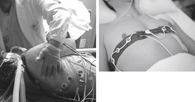 Left: early setup with separate electrodes fixed to the patient. Right: the
individual electrodes are replaced by an electrode belt [6]
Clinicians can follow the regional ventilation of the lungs directly from the bedside screen, which also gives a good sense of the spatial resolution in practice.
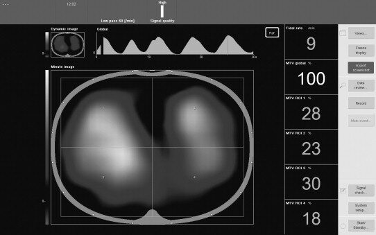 Bedside screen where the patient’s lung images can be followed continuously
[5]
EIT is also used to monitor lung ventilation in newborns. Because it is non-invasive and involves no ionizing radiation, it is particularly suitable for fragile patients such as preterm infants.
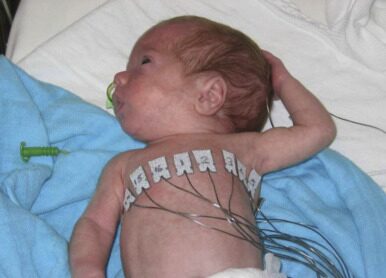 Electrode placement for monitoring lung volume changes in a preterm infant [6]
Breast Imaging
In 2014, a group in Malaysia evaluated an EIT based breast imaging device for cancer screening on 150 women over 40 years old [7]. The device uses a 6 cm x 6 cm planar array with 256 electrodes and a 50 kHz excitation current.
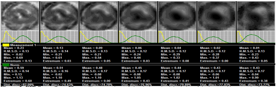 Seven breast slices captured with the EIT device. The second row shows the
conductivity distribution of each slice [7]
Tomo: Gesture Recognition with EIT
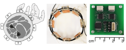
Tomo is a project from Carnegie Mellon University that uses EIT to recognize hand gestures with a small wristband [8]. The band has 8 electrodes, runs at a 40 kHz excitation frequency, and is built from low cost parts. The cross-sectional impedance images of the wrist are fed into a machine learning classifier that predicts the current hand gesture.
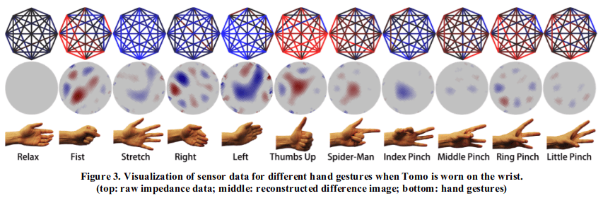 Reconstructed cross sections of the wrist for different hand gestures [8]
The confusion matrices below show the classification accuracy when the band is worn on the wrist and on the arm. One of the key issues the authors mention is electrode contact: the electrodes need to sit tightly on the skin, and a poor contact quickly hurts classification accuracy.
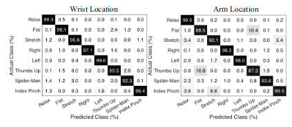 Confusion matrices for the hand gesture set when Tomo is worn on the wrist
(top) and on the arm (bottom) [8]
Spectra: Open Source Biomedical Imaging
Spectra, by Mindseye Biomedical, is an open source project that aims to make biomedical imaging accessible to everyone [9]. Its EIT board is meant for both hardware and algorithm research. It costs around 400 dollars, supports up to 32 electrodes, has an adjustable excitation frequency from 1 kHz to 160 kHz, and comes with a web interface for testing reconstruction algorithms in real time.
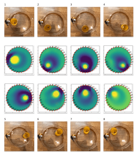
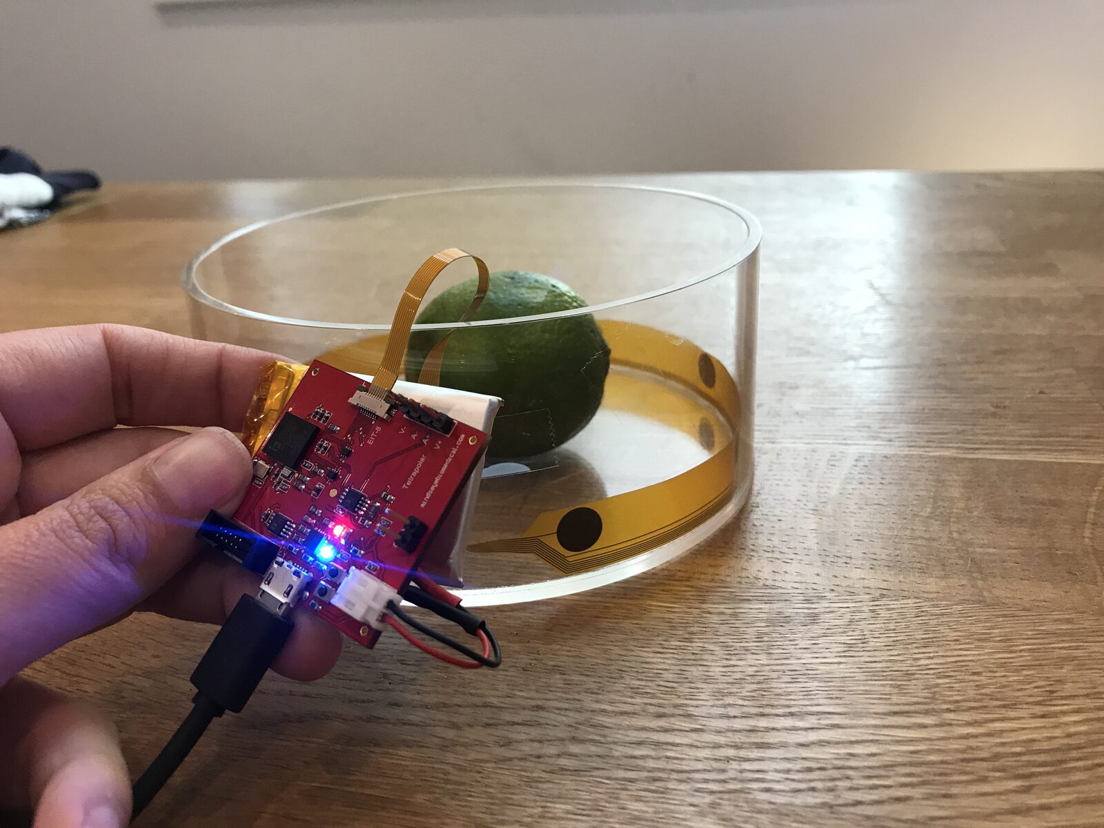 Spectra development board while testing with an EIT phantom [9]
The board has a small form factor and can be tested directly on a phantom through a circular electrode array with a ready to use connector.
Open Problems and Future Directions
EIT is already useful in practice, but there is still a lot of room for improvement.
Physical setup
- Water bath based solutions
- Higher electrode counts
- The skin to electrode interface and its capacitance
Spatial resolution
- Better methods for the ill posed inverse problem
- Modelling 3D current paths instead of assuming 2D slices
- Multi-frequency impedance measurements
- Combining EIT with other imaging modalities
Conclusion
EIT is a young imaging modality with a lot of potential. Its low cost and portability set it apart from the other modalities, and it is especially attractive when tissue needs to be imaged continuously for long periods. I expect it to find many new applications in the coming years, and maybe one day it will help make biomedical imaging accessible to people all around the world.
References
- J. L. Oschman, “Chapter 6: Electricity and Magnetism in Diagnosis and Therapeutics,” in Energy Medicine, 2nd ed., Churchill Livingstone, 2016, pp. 75 to 99.
- L. Borcea, “Electrical Impedance Tomography,” Inverse Problems, 2002.
- J. Malmivuo and R. Plonsey, Bioelectromagnetism, Chapter 26: Impedance Tomography, Oxford University Press, 1995.
- K. Aristovich, G. Sato dos Santos, B. Packham, and D. Holder, “A method for reconstructing tomographic images of evoked neural activity with electrical impedance tomography using intracranial planar arrays,” Physiological Measurement, vol. 35, p. 1095, 2014.
- S. Grimnes and Ø. G. Martinsen, “Chapter 10: Selected Applications,” in Bioimpedance and Bioelectricity Basics, 3rd ed., Academic Press, 2015.
- J. Stocks and S. Lum, “Pulmonary Function Tests in Infants and Preschool Children,” in Kendig and Chernick’s Disorders of the Respiratory Tract in Children, 8th ed., W.B. Saunders, 2012.
- N. Mohd Zain and K. Chelliah, “Breast Imaging Using Electrical Impedance Tomography: Correlation of Quantitative Assessment with Visual Interpretation,” Asian Pacific Journal of Cancer Prevention, vol. 15, pp. 1327 to 1331, 2014.
- Y. Zhang and C. Harrison, “Tomo: Wearable, Low-Cost Electrical Impedance Tomography for Hand Gesture Recognition,” in Proceedings of UIST, 2015.
- J. Rintoul, “Mindseye Biomedical Spectra: Open Source Biomedical Imaging,” Crowd Supply, 2019. https://www.crowdsupply.com/mindseye-biomedical/spectra
← Playing DOTA on Raspberry Pi using Steam Link | Human Activity Recognition with LSTM Networks →